PROBE
provides extensive tools
for rendering, enhancing, analyzing
and annotating 3D rendered images,
some of which are described here:
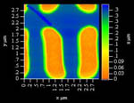
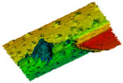
| Dimensional Integrity : |
|
Dimensional axes may be shown or, as in this illustration, the axes may be embedded in the image. Dimensional integrity is maintained regardless of modifications to the image. |
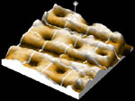
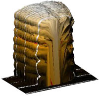 Side wall profiling AFM IBM |
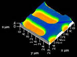
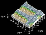
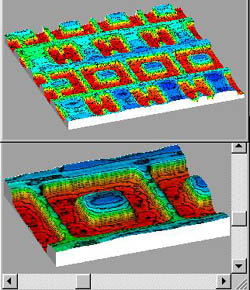
| Presentation Modes: |
|
3D rendered images may be shown in any of four presentation modes. Even in the 2D mode, Z-axis information is retained and shown.
|
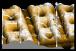
Non-contact
AFM |
|
|
Four image presentation modes are shown. A 3D image in any presentation mode can be rotated a full 360 degrees about both the X and the Y axes.
|
|
Selected
areas of interest within images can be zoomed to enlarge features.
The adjustable zoom window can be moved within the full image.
|
|
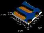
| Color Mapping |
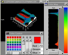 Non-Contact AFM |
Images may be color mapped in several modes. Shown is Spectral mode, using multiple colors to indicate Z dimensions. The image can also be color mapped in Topographic mode, where color bands are delineated, as in a contour map. Gradation maps a single color, using brightness to indicate Z. |
|
Range of color (or brightness) can be selected and applied to a selected range of the Z data histogram. Relative Z dimensions may be color coded by color mapping. |
 Confocal Optical Imaging Damaged mask |
| Filtering |
|
Extensive filtering facilities are available to accentuate or enhance selected features or to reduce noise. Illustrated is a 2DFFT enhancement of a section of the upper edge of an etched hole. |
Other available filtering tools include: Smoothing, Sharpening, LaPlace, Median, Least Squares, Sobel, Mean Value, Three-Point Leveling, plus a 5 X 5 user-definable filter matrix. |
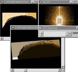
 Interferometric microscope Shallow sub-micron structure in silicon |
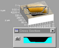
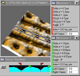
Cross sectional slice showing profile. |
| Clicking within the profile window activates measuring tools. Extensive measurement capabilities are provided, using the ruler tools. Up to four Measurement Tables can be created from a single cross sectional window. |
Each table includes d(x), d(y), d(z), d(xy), d(xyz) and the angle between any two se-lected points. |
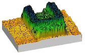
| 3D Renderings from Optical Images: |
Stacks of 2D images can be rendered as 3D volumes (left) and 3D height maps (right). Cross sectional tool shows relative elevations.
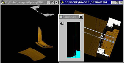
Optical
microscope imaging
Intersecting channels
| Interferometric Images: |
PROBE can import image files from a wide variety of imaging sources, plus Excel and MathCad files. Probe can accept interferomic and SPM image data from Veeco, and other interference and SPM microscope manufacturers, and from specialty imaging systems such as FRT. |
 |
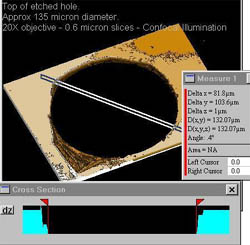 Optical microscope imaging Top 10 microns of damaged etched hole 135 micron dia. |
The Select and Draw tools provide a full capability to add data in text and graphics to the images developed with Probe. Using the SuperClip module, composite images such as this can be captured and saved in all standard graphic file formats. |
| Locating
Tools : |
|
This group of tools provides extensive capabilities for identifying objects for further processing. |
| Level
Setting Tool: |
|
This tool allows setting profile level to accentuate features. |
Probe includes many additional enhancement, analysis and annotation tools. To download a demonstration version of the software, click Download.
Features
![]()
![]()
ABOUT | PROBE 3D | SMARTFOCUS | GALLERY | DEMO | CONTACT | HOME
COPYRIGHT
2001- GENERAL NANOTECHNOLOGY. LLC. ALL RIGHTS RESERVED.